Review

Importance of Cry Proteins in Biotechnology: Initially a Bioinsecticide, Now a Vaccine Adjuvant

by
Maria Cristina Gonzalez-Vazquez
1,
Ruth Abril Vela-Sanchez
2,
Norma Elena Rojas-Ruiz
1,2 and
Alejandro Carabarin-Lima
1,2,*
1
Centro de Investigaciones en Ciencias Microbiologicas, Instituto de Ciencias, Benemerita Universidad Autonoma de Puebla, Puebla 72000, PU, Mexico
2
Licenciatura en Biotecnología, Benemerita Universidad Autonoma de Puebla, Puebla 72000, PU, Mexico
*
Author to whom correspondence should be addressed.
Life 2021, 11(10), 999; https://doi.org/10.3390/life11100999
Submission received: 7 September 2021 / Revised: 19 September 2021 / Accepted: 19 September 2021 / Published: 23 September 2021
(This article belongs to the Section Medical Research)

Abstract

:
A hallmark of Bacillus thuringiensis bacteria is the formation of one or more parasporal crystal (Cry) proteins during sporulation. The toxicity of these proteins is highly specific to insect larvae, exerting lethal effects in different insect species but not in humans or other mammals. The aim of this review is to summarize previous findings on Bacillus thuringiensis, including the characteristics of the bacterium, its subsequent contribution to biotechnology as a bioinsecticide due to the presence of Cry proteins, and its potential application as an adjuvant. In several studies, Cry proteins have been administered together with specific antigens to immunize experimental animal models. The results have shown that these proteins can enhance immunogenicity by generating an adequate immune response capable of protecting the model against an experimental infectious challenge, whereas protection is decreased when the specific antigen is administered without the Cry protein. Therefore, based on previous results and the structural homology between Cry proteins, these molecules have arisen as potential adjuvants in the development of vaccines for both animals and humans. Finally, a model of the interaction of Cry proteins with different components of the immune response is proposed.

1. Introduction

The aim of this review is to show the potential use of Cry proteins as vaccine adjuvants. First, general information is presented about Bacillus thuringiensis (Bt), a bacterium that can form spores and produce Cry proteins, which are natural bioinsecticides. Then, the review focuses on a new property attributed to Cry proteins: immunopotentiators of the immune response or adjuvants. This is an innovative application to explore because, currently, few adjuvants are licensed for vaccine formulations, and several are in the research stage [1]. Additionally, identifying a natural adjuvant that does not cause harm to humans will be beneficial, and Cry proteins are optimal candidates to play this role in the near future [2,3,4]. Moreover, recent research in animal models has shown that Cry proteins are not toxic [5], and their use in human vaccines could be safe.

2. Bacillus Thuringiensis (Bt)

B. thuringiensis was isolated for the first time in 1902 by the Japanese scientist Ishiwata, who was studying the cause of mortality in silkworm larvae; thus, this disease was also called Soto disease. Ishiwata initially named this bacterium Bacillus sotto [6]. A few years later, in 1911, a German scientist, Ernst Berliner, isolated a bacterial strain in dead moth larvae in Mediterranean flour, located in a flour mill in the German state of Thuringia. For this reason, Ernst named this Bacillus B. thuringiensis (Bt) [7]. Subsequently, the probable mechanism of cytotoxic action of particular Bt inclusions, called parasporal, was shown in silkworm larvae (Bombyx mori). Changes in the permeability of the intestinal walls of the insect were observed, consequently causing its death. These results showed that the parasporal inclusions contained crystals of δ-endotoxin, which was the cause of the larva deaths [8,9]. The successful use of Bt in agriculture lies in the production of crystal proteins called Cry, which have specific cytotoxic activity against different insect orders, such as Lepidoptera, Diptera, Coleoptera, Hymenoptera, Homoptera, Orthoptera, and Mallophaga [10,11,12].
The first product based on Bt toxins was commercialized in 1938 in France for moth control. In 1958, Bt products became commercially available in the USA [13]. The predominant use of Bt toxins is for the control of agricultural pests, which is carried out by genetically modified plants (Bt plants) [14,15,16,17]. Several excellent reviews have been published on the control of pests using Cry toxins [18,19,20,21]. Table 1 shows some of the pests controlled by Cry toxins.

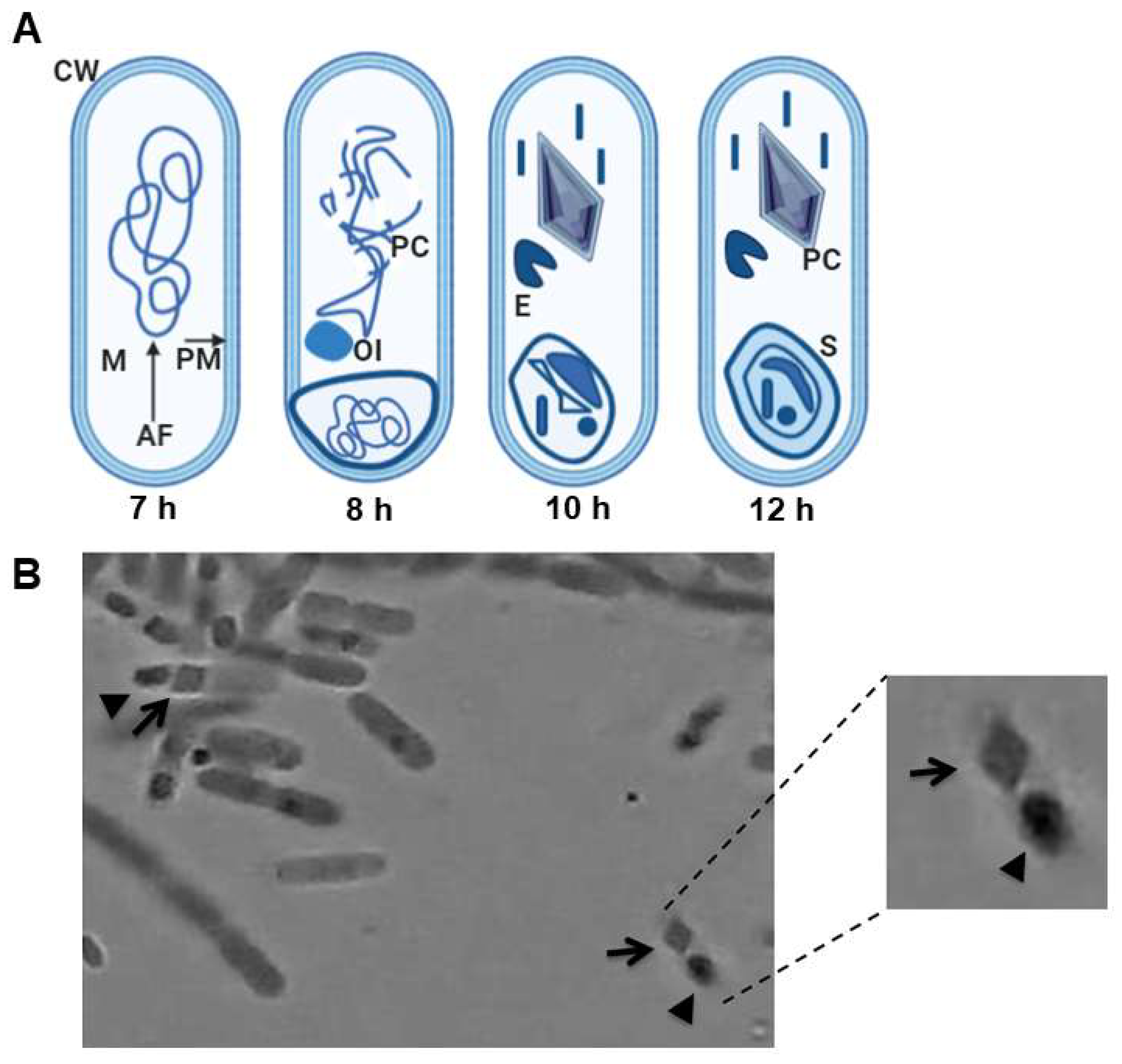
2.1. Overview of Bt and Spore Formation

Bt is a Gram-positive, catalase-positive, oxidase-negative, strictly aerobic bacterium with peritrichous flagella that enable motility. It is generally found in soils, but it is also present in water, insects, some grains, and the environment [11,22,23,24,25]. Bt has two life cycles: vegetative growth and a sporulation phase. One of the main characteristics of Bt that distinguishes it from other bacilli of the same genus is the intracellular presence of a protein crystal [11,15,17,26].
During the sporulation phase, Bt forms a protein crystal. The development of the spore and crystal comprises seven distinct stages: (a) phase I: axial filament formation; (b) phase II: formation of the forespore septum; (c) phase III: parasporal crystals and pre-spore formation; (d) phases IV–VI: exospore formation, primordial cell wall development, and the transformation of spore nucleoids; (e) phase VII: spore maturation and cell lysis (Figure 1) [6,10,27,28]. Crystals are synthesized after stage II of sporulation and accumulate in the cell where they can represent up to 30% of the dry weight of the sporulated cells [29,30]. Several authors have described the presence of different forms of Bt crystals, such as dipyramidal, pyramidal, cuboidal, flat rhomboid, spherical, and rectangular. The most commonly found shape is the dipyramidal crystal. These crystals may include one or more δ-endotoxins, also known as Cry proteins [31,32,33].
In 1981, the cry gene that encodes for the toxin protein in Bt was cloned for the first time [34]. To date, 731 genes encoding Cry proteins have been found with 272 holotypes [35]. Cry proteins are classified into 50 groups and several subgroups, depending on the host specificity, structure, and mechanism of action. These proteins have molecular weights between 30 kDa and 140 kDa [36,37].

2.2. Cry Proteins

In 1989, a nomenclature was proposed to classify proteins according to their sequence and specificity. In this initial nomenclature, there were only four classes. The first class included proteins with action against Lepidoptera with a size of approximately 130–140 kDa. The second class included smaller proteins (65 kDa) with activity against Lepidoptera and Diptera; this class included only two members: CryIIA and CryIIB. The third class constituted the active toxin against Coleoptera, CryIIIA. The last class was Cry1A, the members of which were closely related: they were called Cry1Aa, Cry1Ab, and Cry1Ac [14].
In 1998, a new nomenclature was published classifying toxins solely by their amino acid sequence. On this basis, most proteins are related and contain up to five conserved domains. Subsequently, a slightly modified name system was adopted in which each toxin receives a name that incorporates four levels. First, in general, toxins that share at least 45% identity in their sequence have the same number. The second rank (A) is used to distinguish sequences that share between 45% and 78% identity. Those that share between 78% and 95% identity are distinguished at the tertiary level (a). Finally, the quaternary range is used to identify certain differences. Subsequently, in 2003, Cry proteins were classified into three groups through a phylogenetic approach. The group with the majority of Cry toxins is known as the family of three domains since they contain three structural domains. This family contains the largest group of Cry proteins, which are globular molecules that contain three structural domains connected by simple bonds. A particular characteristic of the members of this family is the presence of protoxins with two different lengths. Long protoxins are approximately twice the length of most toxins. The C-terminal extension found in long protoxins is dispensable for toxicity and is believed to play a role in the formation of crystal inclusion bodies within the bacteria [14]. To date, the three dimensional structures of 12 Cry toxins without modifications (Cry1Aa6, Cry1Ac7, Cry1Ac8, Cry2Aa, Cry3Aa12, Cry3Aa3, Cry3Bb1, Cry4Aa1, Cry4Ba1, Cry5Ba1, Cry7Ca1, and Cry8Ea1) [38,39,40,41,42,43,44,45,46,47,48], and some with modifications in the form of mutations (Cry1Ac4 and Cry1Da1) [49,50], have been determined by X-ray crystallography (Figure 2).
The tertiary structure of the N-terminal domain, called domain I, is a set of seven α-helices, among which the central α-helix is hydrophobic and surrounded by six amphipathic helices; this helical domain is responsible for membrane insertion and pore formation. Domain II consists of three antiparallel β sheets with exposed loop regions, and domain III is a β sheet [38,39,40,41,42,46]. The most exposed regions in the tertiary structure of the protein are domains II and III, which are involved in receptor binding [50].
The crystal protein consists of proteins called δ-endotoxin. The definition of Cry proteins is any parasporal protein of Bt that shows a toxic effect on an organism, verifiable using bioassays, or any protein that shows similarity to Cry proteins [51].

2.3. Action Mechanism of Cry as a Bioinsecticide

Crystal proteins have been widely used in genetically modified cultures; these transgenic cultures can produce Bt crystals, making them insect resistant. The two presentations of bioinsecticides, those that contain spores and toxic crystals or transgenic foods that express Cry proteins, have a similar mechanism [52]. Cry protein crystals need to be solubilized for their activation after they are ingested by an insect larva. The activation is mediated by the action of proteolytic enzymes, such as cathepsin G and chymotrypsin, which are located in the digestive system of the insect larva [53] and perform proteolytic processing at the amino terminus of the Cry protein [54]. Once the protein is activated it becomes the so-called δ-endotoxin; this soluble and partially truncated form of the protein is expressed in transgenic foods [52]. The δ-endotoxin binds to receptors located in the membrane of the epithelial cells of the intestine of the larva. Proteins such as cadherins [55], aminopeptidases, and alkaline peptidases [56] are recognized by the domains of δ-endotoxin and, recently, the binding of δ-endotoxin to other proteins, such as ATP-binding transporter proteins (ABC), has been demonstrated [57]. The cadherins that bind to Cry toxins in different orders of insects share a structure composed of four domains: an ectodomain (CE), a domain of the proximal extracellular membrane (MPED), a transmembrane domain (TM), and a cytoplasmic domain (CYTO). Several models have been proposed to explain the action mode of Cry proteins. One of them describes the process in several steps: solubilization of the crystal, processing of the protoxins, and binding to the receptor. The binding allows the oligomerization of δ-endotoxin in the membrane of intestinal cells, its insertion into the membrane, and aggregation, which results in the formation of a pore. This pore in the cell membrane causes an ionic imbalance (release of H+, K+, Na+, and Ca2+ ions) in the cell [58,59,60], which causes an increase in cAMP and, consequently, the activation of the apoptotic process known as cytolysis (Figure 3) [61]. Another model suggests that the toxin monomer can bind to a cadherin receptor and activate Mg2+-dependent signal transduction, a pathway that leads to cell death [62].
Some non-target insects and even some mammals, such as humans, are not sensitive to this bioinsecticide despite having the same receptors on the cell membrane; however, a difference in the structures of the receptors has been observed. Cadherin (Type IV) proteins in sensitive insects have eight or more cadherin domains, which facilitate the anchoring of δ-endotoxin, unlike the cadherins of resistant insects, which have few domains. For this reason, δ-endotoxin is specific because it binds to certain receptors in target insects [63,64]. Moreover, the proteins that allow proteolytic processing for the activation of the Cry protein are not present in the digestive system of resistant insects [54].
On the other hand, cadherins (type I) in humans have structural differences compared to insect cadherins; they principally have minor ectodomains (EC) and a few Ca2+ insertions, which confer foldability to the consecutive extracellular cadherin domains responsible for homophilic binding. This binding is also different in human cadherins because the EC1 domain of vertebrate cadherins contains a conserved tryptophan residue (W) inserted in the hydrophobic pocket, affecting homophilic binding [65]. Moreover, the identity between the cadherins of humans and those of insects (Diptera, Lepidoptera, and Coleoptera) is very low, ranging from 13% to 20% (Table 2). For these reasons, Cry proteins do not pose any potential toxicological risk to humans when they are ingested.

3. Adjuvants Currently in Use

Traditional vaccines derived from toxins, or attenuated or inactivated live organisms are effective in inducing predominantly antibody-based immunity. Adjuvants (taken from Latin “adjuvare”, meaning “help”) are designed to improve the immune response to the vaccine [66]. Adjuvants were initially described as “substances used in combination with a specific antigen that produces a more effective immune response than the antigen alone”, thus encompassing a wide range of substances that can potentially function as an adjuvant [67].
Gaston Ramon, a French veterinarian, observed that the performance of antisera against tetanus and diphtheria in horses was higher in animals that developed an abscess at the injection site. By injecting starch, breadcrumbs, or tapioca, abscesses were sterilized at the site of injection and were, therefore, able to increase the production of antisera [68]. Subsequently, aluminum was used as an adjuvant in vaccines in 1932 [69]. Direct immunization with most antigens will lead to a poor immune response and rapid elimination of the antigen from the body. To avoid this, the antigen is first combined with an adjuvant. The formation of a stable emulsion between the antigen and the adjuvant allows the sustained presentation of the antigen to the immune system, and the removal of the antigen from the body is delayed. These agents can accelerate the development of the immune response against the antigen and ultimately lead to a stronger adaptive response than would otherwise be possible. The adjuvant should also provide the additional benefit of allowing the use of smaller amounts of the antigen [70]. Some important characteristics required of adjuvants are a high stability, a long useful life, biodegradability, and low cost to produce or obtain, and they should not induce immune responses against themselves but induce an appropriate immune response according to the requirements (immune response mediated by antibodies or cells) [66].
Many molecules have been considered for their use as adjuvants. Adjuvants have been classified into two groups: those that direct the antigen to antigen-presenting cells (APCs) and immunostimulatory types, which directly activate cells through specific receptors, producing inflammatory responses that amplify the innate immune response [69]. The ultimate aim of using an adjuvant is to activate the innate immune system to respond more quickly to stimuli and to enhance the specificity of the adaptive immune response [71]. Despite the multiple advantages of adjuvants for the potential action of a vaccine, only a few are currently used in licensed human vaccines (Table 3).

3.1. Benefits of Cry Relative to Other Adjuvants

Adjuvants that are currently used in human vaccines exhibit diverse immune response mechanisms. One of the well-characterized adjuvants is MF59. This adjuvant does not generate Th1-type immunity and, thus, its use in vaccines that are required to induce cell-mediated immunity for protection is not possible [84]. Another adjuvant is Lipopolysaccharide (LPS), which can induce the production of cytokines and chemokines in a variety of cells that can control the traffic and maturation of DC [85]. However, LPS has limitations that derive from its pyrogenicity and toxicity in animals and humans, so its use in vaccines as an adjuvant is rare.
Regarding the study of new adjuvants, cholera toxin (CT) from Vibrio cholerae and heat-labile toxin (LT) from Escherichia coli have been studied as mucosal adjuvants. Unfortunately, both are products derived from bacteria that are pathogenic to humans, preventing their use in vaccines despite being very good activators of the mucosal immune response. Furthermore, they have high production costs [86,87,88].
In this context, the emerging potential of Cry proteins as adjuvants is important because they are not toxic to vertebrates, including humans, and the cost of production is relatively low [26,89]. Moreover, their immunogenic and adjuvant capabilities, which are as potent as those of cholera toxin, have already been demonstrated [90].
Among the mechanisms of immune activation, the importance of specific antigen-enhancing adjuvants has been shown in various studies, which have demonstrated that their administration can activate both humoral and cellular immune responses [90,91,92,93]. The route of administration is also very important. Studies have shown that when administered orally, the Cry protein is highly immunogenic, requiring a very low dose, so the possible toxicity of this protein would be greatly diminished [5].
Because of these broad characteristics, Cry proteins are important proteins for the development of adjuvants for vaccine formulations against intra- or extracellular microorganisms. As previously discussed, adjuvants approved for vaccines are limited, and some of them only induce an immune response mechanism or are toxic in high doses.

3.2. An Overview of Cry as a Natural Adjuvant

A strategy used in the design of vaccines to enhance their immunogenicity includes the co-administration of adjuvants that stimulate and improve immunity. Cry proteins have been described as possible adjuvants for their resistance and stability in highly alkaline environments. The structural characteristics of proteins allow them to modulate the immune response and function as adjuvants. For this reason, several proteins have been studied in the context of therapeutic proteins [94]. Importantly, a wide variety of proteins have been used as adjuvants to immunize animals intranasally, which can stimulate a protective immune response in the lungs and upper respiratory tract and possibly at distant sites, such as the gastric and genital mucosa [95].
With respect to mucosal vaccines, when rhesus macaques were vaccinated intranasally with a trimeric gp41 protein coupled to virosomes to evaluate an HIV-1 vaccine, IgA antibodies increased in the genital tract, and immunization also prevented transmission of infection [96]. Studies have been conducted to identify proteins able to induce a mucosal immune response, which was initially realized by Guimares et al. when they demonstrated the immunogenicity of the Cry1Ab protein of Bacillus thuringiensis. When Cry1Ab was exposed to different pH values, the results showed that the protein was only slightly degraded at pH 2.0 and, most importantly, it maintained its immunoreactivity [97]. Subsequently, Cry proteins were used as adjuvants for their resistance and stability. This was demonstrated by immunizing BALB/c mice intranasally, which is an alkaline environment. After immunization, the mice were protected when they were infected by the bacterium Streptococcus pneumoniae [93] or the parasite Naegleria fowleri [98].
Because of these characteristics, several research groups have studied the use of Cry as a potential adjuvant [93,98,99,100]. The role of Cry proteins as vaccine adjuvants was initially observed with the Cry1Ac protein, which was administered by different routes, including intragastric, intraperitoneal (i.p.), and intranasal immunization [92]. The last two are the most efficient due to their ability to induce isotypes of IgA and IgG antibodies in the murine model. These antibodies demonstrated protection in animal models when they were infected with the bacterium Brucella abortus or with parasites Naegleria fowleri, Plasmodium chabaudi, Plasmodium berghei, and Taenia crassiceps [92,98,101,102,103]. In the case of B. abortus, an intracellular bacterium, the use of Cry1Ac with the RB51 B. abortus strain conferred protection against an intranasal challenge with the virulent strain B. abortus 2308 in BALB/c mice. The results showed that the vaccine conferred immunoprotection, as evidenced by a decrease in the splenic bacterial load in immunized animals. The proliferation of cytotoxic TCD8+ cells increased the production of TNF-α and IFN-γ, and the generation of an IgG2a antibody response was also observed. These results indicate that the use of the Cry1Ac protein as a mucosal adjuvant via the intranasal route may be a promising strategy for developing a vaccine against brucellosis [102].
When the Cry1Ac protein was administered together with total extracts of the free-living amoeba N. fowleri, the immunized animals had 100% protection against the development of meningoencephalitis; however, when the animals were immunized with the Cry1Ac protein alone, only 60% of infected mice survived [98].
On the other hand, mice previously treated with Cry1Ac and infected with P. chabaudi (considered non-lethal) had 100% survival compared to mice previously treated with PBS, which demonstrated 80% survival. Furthermore, mice previously treated with Cry1Ac and subsequently infected with P. berghei (lethal parasite) survived longer (12 days) than control mice previously treated with PBS, which died on day 9 post-infection. Regarding the induced immune response, an increase in IFN-γ and TGF-β cytokines was demonstrated, in addition to an increase in the levels of IgG and IgM immunoglobulins, in animals treated and infected with two types of Plasmodium [103].
Conversely, when mice were immunized with the Cry1Ac protein and total lysates of T. crassiceps, only 40% protection was observed in the experimentally infected animal model, and mice immunized only with Cry1Ac did not survive [92,98,99,100,101]. These results demonstrate that Cry1Ac alone is not able to generate a specific immune response; however, when it is used in the company of a specific antigen, it potentiates the immune response and can function as an adjuvant. At present, the immunological mechanism underlying the effects of the Cry1Ac protein is not entirely understood, but in macrophages this protein can stimulate the overexpression of surface glycoproteins CD80 and CD86, which stimulate the secretion of TNF-α and IL-6 cytokines, allowing activation of an immune response that promotes the survival of animal models against some experimental infections [104].
Another protein used as a potential adjuvant is Cry1Ab (a protein with 86% homology at the amino acid level with Cry1Ac). When administered intranasally, this protein was not able to generate serum and mucosal IgG antibody responses. It was only able to elicit a low level of IgM and SIgA. In contrast, when Cry1Ab was administered by the intraperitoneal route, it was able to induce high levels of IgG and IgM antibodies, similar to the effects of Cry1Aa and Cry1Ac [105]. In another study, the administration of Cry1Ab (1 µg) to BALB/c mice by the i.p. route induced a mixed Th1-Th2 immune response. No evidence of allergenicity has been observed with the administration of Cry proteins [106], though the levels of leukotrienes, cytokines, and eosinophils have notably increased. However, another study conducted with the direct consumption of Bt corn with Cry1Ab protein showed that the inclusion of Cry1Ab in the diet did not affect the severity of asthma or allergic inflammation induced by ovalbumin [104].
Based on the previously described research, the possible immunological mechanism of Cry proteins may be the following: Cry proteins may be recognized by a Toll-like receptor located on the surface of antigen-presenting cells such as macrophages; many current adjuvants are recognized by various members of the Toll-like receptor (TLR) family [107]. These activated macrophages can secrete the IL-8 cytokine into the medium, which will recruit inflammatory cells such as neutrophils and immunological B and T lymphocytes. Once the immunodominant epitopes are exposed in the major histocompatibility complex type 1, they will be recognized by the TCR receptor of CD4+ lymphocytes, which will secrete cytokines such as IL-6, IL-8, TNF-α, and INF-γ. This last cytokine will be recognized by the INF-γ receptor exposed on B lymphocytes. Once the cytokine binds to the receptor, a series of signaling pathways will be activated inside the lymphocytes, which will induce the secretion of IgG2a and IgG2b antibodies (Figure 4).
The profile of cytokines and immunoglobulins activated by Cry proteins is the Th1 type; therefore, CD4+ T lymphocytes that differentiate into a subpopulation of T helper 1 cells could also activate this immune response. This type of response has been described as ideal for the elimination of a large variety of bacteria and parasites, principally intracellular. For this reason, a promising approach is to generate fused proteins by exploring Cry proteins together with any important protein of a pathogenic microorganism to use it as an adjuvant and potentiate a suitable immune response.
Finally, the activity of Cry proteins as bioinsecticides has been widely studied [51]; however, their other activities, such as their potential adjuvant function, have been inadequately explored. Although some experiments have confirmed their potential action as immunopotentiators [92,99,101], there are many Cry proteins that have not been evaluated yet. Furthermore, in different studies, the lack of toxicity of these proteins has been demonstrated in mammals, especially humans, and it has even been shown that these proteins can generate an immune response mediated by antibodies [108]. However, it is crucial to determine the mechanism by which Cry proteins are activated to induce an immune response, as well as to evaluate the possible risks that they could pose in the short and long term, such as allergies or other immune alterations. Therefore, we conclude that the use of Cry proteins as natural adjuvants is an interesting opportunity for new biotechnological processes and vaccine development.

4. Conclusions

To protect the health of the population against emerging and re-emerging infectious agents, the development of effective strategies is crucial, including vaccination. The current worldwide pandemic caused by the SARS-CoV-2 virus led to the rapid development of effective vaccines, which was possible because their platforms (adenovirus and mRNA) and adjuvants had been previously shown to work successfully. Requirements for adjuvants include low-cost components, a high production capacity, safety in humans, and the adequate potentiation of the immune response to the vaccine antigen. Some experimental studies have shown that Cry proteins from Bacillus thuringiensis are among such adjuvants. Therefore, in the coming years, it is important to study Cry proteins as adjuvants and begin to use them to promote the development of vaccines against new and old infectious agents.

Author Contributions

Conceptualization, A.C.-L. and M.C.G.-V.; methodology, R.A.V.-S.; software, A.C.-L.; validation, M.C.G.-V. and N.E.R.-R.; formal analysis, R.A.V.-S. and N.E.R.-R.; investigation, M.C.G.-V., R.A.V.-S., and N.E.R.-R.; writing—original draft preparation, R.A.V.-S., N.E.R.-R., and M.C.G.-V.; writing—review and editing, A.C.-L. and M.C.G.-V.; visualization, R.A.V.-S.; supervision, N.E.R.-R.; project administration, A.C.-L. All authors have read and agreed to the published version of the manuscript.

Funding

This research received no external funding.

Institutional Review Board Statement

Not applicable.

Informed Consent Statement

Not applicable.

Data Availability Statement

Not applicable.

Conflicts of Interest

The authors declare no conflict of interest.

References

  1. Apostolico Jde, S.; Lunardelli, V.A.; Coirada, F.C.; Boscardin, S.B.; Rosa, D.S. Adjuvants: Classification, Modus Operandi, and Licensing. J. Immunol. Res. 2016, 2016, 1459394. [Google Scholar] [CrossRef] [PubMed] [Green Version]
  2. Garcon, N.; Di Pasquale, A. From discovery to licensure, the Adjuvant System story. Hum. Vaccin. Immunother. 2017, 13, 19–33. [Google Scholar] [CrossRef]
  3. Sjoblad, R.D.; McClintock, J.T.; Engler, R. Toxicological considerations for protein components of biological pesticide products. Regul. Toxicol. Pharmacol. 1992, 15, 3–9. [Google Scholar] [CrossRef] [Green Version]
  4. Koch, M.S.; Ward, J.M.; Levine, S.L.; Baum, J.A.; Vicini, J.L.; Hammond, B.G. The food and environmental safety of Bt crops. Front. Plant. Sci. 2015, 6, 283. [Google Scholar] [CrossRef]
  5. Mezzomo, B.P.; Miranda-Vilela, A.L.; Grisolia, C.K. Toxicological Evaluation of a Potential Immunosensitizer for Use as a Mucosal Adjuvant--Bacillus thuringiensis Cry1Ac Spore-Crystals: A Possible Inverse Agonist that Deserves Further Investigation. Toxins 2015, 7, 5348–5358. [Google Scholar] [CrossRef] [PubMed] [Green Version]
  6. Ibrahim, M.A.; Griko, N.; Junker, M.; Bulla, L.A. Bacillus thuringiensis: A genomics and proteomics perspective. Bioeng. Bugs 2010, 1, 31–50. [Google Scholar] [CrossRef] [PubMed] [Green Version]
  7. de Barjac, H.; Bonnefoi, A. A classification of strains of Bacillus thuringiensis Berliner with a key to their differentiation. J. Invertebr. Pathol. 1968, 11, 335–347. [Google Scholar] [CrossRef]
  8. Fast, P.G.; Angus, T.A. Effects of Parasporal Inclusions of Bacillus Thuringiensis Var. Sotto Ishiwata on the Permeability of the Gut Wall of Bompyx Mori (Linnaeus) Larvae. J. Invertebr. Pathol. 1965, 20, 29–32. [Google Scholar] [CrossRef]
  9. Fast, P.G.; Donaghue, T.P. The delta-endotoxin of Bacillus thuringiensis. II. On the mode of action. J. Invertebr. Pathol. 1971, 18, 135–138. [Google Scholar] [CrossRef]
  10. Jouzani, G.S.; Valijanian, E.; Sharafi, R. Bacillus thuringiensis: A successful insecticide with new environmental features and tidings. Appl. Microbiol. Biotechnol. 2017, 101, 2691–2711. [Google Scholar] [CrossRef]
  11. Schnepf, E.; Crickmore, N.; Van Rie, J.; Lereclus, D.; Baum, J.; Feitelson, J.; Zeigler, D.R.; Dean, D.H. Bacillus thuringiensis and its pesticidal crystal proteins. Microbiol. Mol. Biol. Rev. 1998, 62, 775–806. [Google Scholar] [CrossRef] [PubMed] [Green Version]
  12. Wei, J.Z.; Hale, K.; Carta, L.; Platzer, E.; Wong, C.; Fang, S.C.; Aroian, R.V. Bacillus thuringiensis crystal proteins that target nematodes. Proc. Natl. Acad. Sci. USA 2003, 100, 2760–2765. [Google Scholar] [CrossRef] [Green Version]
  13. Lambert, B.; Hofte, H.; Annys, K.; Jansens, S.; Soetaert, P.; Peferoen, M. Novel Bacillus thuringiensis insecticidal crystal protein with a silent activity against coleopteran larvae. Appl. Environ. Microbiol. 1992, 58, 2536–2542. [Google Scholar] [CrossRef] [PubMed] [Green Version]
  14. Raymond, B. The biology, ecology and taxonomy of Bacillus thuringiensis and related bacteria. In Bacillus thuringiensis and Lysinibacillus sphaericus; Springer: Berlin/Heidelberg, Germany, 2017; pp. 19–39. [Google Scholar]
  15. Hofte, H.; de Greve, H.; Seurinck, J.; Jansens, S.; Mahillon, J.; Ampe, C.; Vandekerckhove, J.; Vanderbruggen, H.; van Montagu, M.; Zabeau, M.; et al. Structural and functional analysis of a cloned delta endotoxin of Bacillus thuringiensis berliner 1715. Eur. J. Biochem. 1986, 161, 273–280. [Google Scholar] [CrossRef] [PubMed]
  16. Key, S.; Ma, J.K.; Drake, P.M. Genetically modified plants and human health. J. R. Soc. Med. 2008, 101, 290–298. [Google Scholar] [CrossRef] [Green Version]
  17. Hofte, H.; Seurinck, J.; Van Houtven, A.; Vaeck, M. Nucleotide sequence of a gene encoding an insecticidal protein of Bacillus thuringiensis var. tenebrionis toxic against Coleoptera. Nucleic. Acids. Res. 1987, 15, 7183. [Google Scholar] [CrossRef] [Green Version]
  18. Chattopadhyay, P.; Banerjee, G. Recent advancement on chemical arsenal of Bt toxin and its application in pest management system in agricultural field. 3 Biotech. 2018, 8, 201. [Google Scholar] [CrossRef]
  19. Christou, P.; Capell, T.; Kohli, A.; Gatehouse, J.A.; Gatehouse, A.M. Recent developments and future prospects in insect pest control in transgenic crops. Trends. Plant. Sci. 2006, 11, 302–308. [Google Scholar] [CrossRef] [PubMed]
  20. Romeis, J.; Meissle, M.; Bigler, F. Transgenic crops expressing Bacillus thuringiensis toxins and biological control. Nat. Biotechnol. 2006, 24, 63–71. [Google Scholar] [CrossRef] [PubMed]
  21. Valtierra-de-Luis, D.; Villanueva, M.; Berry, C.; Caballero, P. Potential for Bacillus thuringiensis and Other Bacterial Toxins as Biological Control Agents to Combat Dipteran Pests of Medical and Agronomic Importance. Toxins 2020, 12, 773. [Google Scholar] [CrossRef]
  22. Bernhard, K. Quantitative determination of delta-endotoxin contents in spray-dried preparations ofBacillus thuringiensis strain GC-91. World J. Microbiol. Biotechnol. 1992, 8, 24–29. [Google Scholar] [CrossRef] [PubMed]
  23. Martin, P.A.; Travers, R.S. Worldwide Abundance and Distribution of Bacillus thuringiensis Isolates. Appl. Environ. Microbiol. 1989, 55, 2437–2442. [Google Scholar] [CrossRef] [Green Version]
  24. Aronson, A.I.; Beckman, W.; Dunn, P. Bacillus thuringiensis and related insect pathogens. Microbiol. Rev. 1986, 50, 1–24. [Google Scholar] [CrossRef] [PubMed]
  25. Ichimatsu, T.; Mizuki, E.; Nishimura, K.; Akao, T.; Saitoh, H.; Higuchi, K.; Ohba, M. Occurrence of Bacillus thuringiensis in fresh waters of Japan. Curr. Microbiol. 2000, 40, 217–220. [Google Scholar] [CrossRef]
  26. Hofte, H.; Whiteley, H.R. Insecticidal crystal proteins of Bacillus thuringiensis. Microbiol. Rev. 1989, 53, 242–255. [Google Scholar] [CrossRef]
  27. Bulla, L.A., Jr.; Bechtel, D.B.; Kramer, K.J.; Shethna, Y.I.; Aronson, A.I.; Fitz-James, P.C. Ultrastructure, physiology, and biochemistry of Bacillus thuringiensis. Crit. Rev. Microbiol. 1980, 8, 147–204. [Google Scholar] [CrossRef] [PubMed]
  28. Bechtel, D.B.; Bulla, L.A., Jr. Ultrastructural analysis of membrane development during Bacillus thuringiensis sporulation. J. Ultrastruct. Res. 1982, 79, 121–132. [Google Scholar] [CrossRef]
  29. Agaisse, H.; Lereclus, D. How does Bacillus thuringiensis produce so much insecticidal crystal protein? J. Bacteriol. 1995, 177, 6027–6032. [Google Scholar] [CrossRef] [Green Version]
  30. Baum, J.A.; Malvar, T. Regulation of insecticidal crystal protein production in Bacillus thuringiensis. Mol. Microbiol. 1995, 18, 1–12. [Google Scholar] [CrossRef]
  31. Lopez-Meza, J.E.; Ibarra, J.E. Characterization of a Novel Strain of Bacillus thuringiensis. Appl. Environ. Microbiol. 1996, 62, 1306–1310. [Google Scholar] [CrossRef] [Green Version]
  32. Fiuza, L.M.; Schunemann, R.; Pinto, L.M.; Zanettini, M.H. Two new Brazilian isolates of Bacillus thuringiensis toxic to Anticarsia gemmatalis (Lepidoptera: Noctuidae). Braz. J. Biol. 2012, 72, 363–369. [Google Scholar] [CrossRef] [Green Version]
  33. Mukhija, B.; Khanna, V. Isolation, characterization and crystal morphology study of Bacillus thuringiensis isolates from soils of Punjab. J. Pure Appl. Microbiol. 2018, 12, 189–194. [Google Scholar] [CrossRef]
  34. Schnepf, H.E.; Whiteley, H.R. Cloning and expression of the Bacillus thuringiensis crystal protein gene in Escherichia coli. Proc. Natl. Acad. Sci. USA 1981, 78, 2893–2897. [Google Scholar] [CrossRef] [Green Version]
  35. Crickmore, N.; Berry, C.; Panneerselvam, S.; Mishra, R.; Connor, T.R.; Bonning, B.C. A structure-based nomenclature for Bacillus thuringiensis and other bacteria-derived pesticidal proteins. J. Invertebr. Pathol. 2020, 107438. [Google Scholar] [CrossRef] [PubMed]
  36. Crickmore, N.; Zeigler, D.R.; Feitelson, J.; Schnepf, E.; Van Rie, J.; Lereclus, D.; Baum, J.; Dean, D.H. Revision of the nomenclature for the Bacillus thuringiensis pesticidal crystal proteins. Microbiol. Mol. Biol. Rev. 1998, 62, 807–813. [Google Scholar] [CrossRef] [Green Version]
  37. Crickmore, N.; Baum, J.; Bravo, A.; Lereclus, D.; Narva, K.; Sampson, K.; Schnepf, E.; Sun, M.; Zeigler, D.R. Bacillus thuringiensis toxin nomenclature. Available online: http://www.btnomenclature.info/ (accessed on 18 September 2021).
  38. Boonserm, P.; Mo, M.; Angsuthanasombat, C.; Lescar, J. Structure of the functional form of the mosquito larvicidal Cry4Aa toxin from Bacillus thuringiensis at a 2.8-angstrom resolution. J. Bacteriol. 2006, 188, 3391–3401. [Google Scholar] [CrossRef] [PubMed] [Green Version]
  39. Li, J.D.; Carroll, J.; Ellar, D.J. Crystal structure of insecticidal delta-endotoxin from Bacillus thuringiensis at 2.5 A resolution. Nature 1991, 353, 815–821. [Google Scholar] [CrossRef] [PubMed]
  40. Grochulski, P.; Masson, L.; Borisova, S.; Pusztai-Carey, M.; Schwartz, J.L.; Brousseau, R.; Cygler, M. Bacillus thuringiensis CryIA(a) insecticidal toxin: Crystal structure and channel formation. J. Mol. Biol. 1995, 254, 447–464. [Google Scholar] [CrossRef]
  41. Galitsky, N.; Cody, V.; Wojtczak, A.; Ghosh, D.; Luft, J.R.; Pangborn, W.; English, L. Structure of the insecticidal bacterial delta-endotoxin Cry3Bb1 of Bacillus thuringiensis. Acta Crystallogr. D Biol. Crystallogr. 2001, 57, 1101–1109. [Google Scholar] [CrossRef] [PubMed] [Green Version]
  42. Boonserm, P.; Davis, P.; Ellar, D.J.; Li, J. Crystal structure of the mosquito-larvicidal toxin Cry4Ba and its biological implications. J. Mol. Biol. 2005, 348, 363–382. [Google Scholar] [CrossRef]
  43. Guo, S.; Ye, S.; Liu, Y.; Wei, L.; Xue, J.; Wu, H.; Song, F.; Zhang, J.; Wu, X.; Huang, D.; et al. Crystal structure of Bacillus thuringiensis Cry8Ea1: An insecticidal toxin toxic to underground pests, the larvae of Holotrichia parallela. J. Struct. Biol. 2009, 168, 259–266. [Google Scholar] [CrossRef] [PubMed]
  44. Hui, F.; Scheib, U.; Hu, Y.; Sommer, R.J.; Aroian, R.V.; Ghosh, P. Structure and glycolipid binding properties of the nematicidal protein Cry5B. Biochemistry 2012, 51, 9911–9921. [Google Scholar] [CrossRef] [PubMed] [Green Version]
  45. Derbyshire, D.J.; Ellar, D.J.; Li, J. Crystallization of the Bacillus thuringiensis toxin Cry1Ac and its complex with the receptor ligand N-acetyl-D-galactosamine. Acta Crystallogr. D Biol. Crystallogr. 2001, 57, 1938–1944. [Google Scholar] [CrossRef] [PubMed] [Green Version]
  46. Morse, R.J.; Yamamoto, T.; Stroud, R.M. Structure of Cry2Aa suggests an unexpected receptor binding epitope. Structure 2001, 9, 409–417. [Google Scholar] [CrossRef]
  47. Sawaya, M.R.; Cascio, D.; Gingery, M.; Rodriguez, J.; Goldschmidt, L.; Colletier, J.P.; Messerschmidt, M.M.; Boutet, S.; Koglin, J.E.; Williams, G.J.; et al. Protein crystal structure obtained at 2.9 A resolution from injecting bacterial cells into an X-ray free-electron laser beam. Proc. Natl. Acad. Sci. USA 2014, 111, 12769–12774. [Google Scholar] [CrossRef] [Green Version]
  48. Jing, X.; Yuan, Y.; Wu, Y.; Wu, D.; Gong, P.; Gao, M. Crystal structure of Bacillus thuringiensis Cry7Ca1 toxin active against Locusta migratoria manilensis. Protein Sci. 2019, 28, 609–619. [Google Scholar] [CrossRef] [Green Version]
  49. Evdokimov, A.G.; Moshiri, F.; Sturman, E.J.; Rydel, T.J.; Zheng, M.; Seale, J.W.; Franklin, S. Structure of the full-length insecticidal protein Cry1Ac reveals intriguing details of toxin packaging into in vivo formed crystals. Protein Sci. 2014, 23, 1491–1497. [Google Scholar] [CrossRef] [Green Version]
  50. Fernandez, L.E.; Perez, C.; Segovia, L.; Rodriguez, M.H.; Gill, S.S.; Bravo, A.; Soberon, M. Cry11Aa toxin from Bacillus thuringiensis binds its receptor in Aedes aegypti mosquito larvae through loop alpha-8 of domain II. FEBS Lett. 2005, 579, 3508–3514. [Google Scholar] [CrossRef] [PubMed] [Green Version]
  51. Palma, L.; Munoz, D.; Berry, C.; Murillo, J.; Caballero, P. Bacillus thuringiensis toxins: An overview of their biocidal activity. Toxins 2014, 6, 3296–3325. [Google Scholar] [CrossRef] [Green Version]
  52. Jurat-Fuentes, J.L.; Crickmore, N. Specificity determinants for Cry insecticidal proteins: Insights from their mode of action. J. Invertebr. Pathol. 2017, 142, 5–10. [Google Scholar] [CrossRef] [Green Version]
  53. Walters, F.S.; Stacy, C.M.; Lee, M.K.; Palekar, N.; Chen, J.S. An engineered chymotrypsin/cathepsin G site in domain I renders Bacillus thuringiensis Cry3A active against Western corn rootworm larvae. Appl. Environ. Microbiol. 2008, 74, 367–374. [Google Scholar] [CrossRef] [Green Version]
  54. Du, C.; Martin, P.A.; Nickerson, K.W. Comparison of Disulfide Contents and Solubility at Alkaline pH of Insecticidal and Noninsecticidal Bacillus thuringiensis Protein Crystals. Appl. Environ. Microbiol. 1994, 60, 3847–3853. [Google Scholar] [CrossRef] [Green Version]
  55. Melo, A.L.; Soccol, V.T.; Soccol, C.R. Bacillus thuringiensis: Mechanism of action, resistance, and new applications: A review. Crit. Rev. Biotechnol. 2016, 36, 317–326. [Google Scholar] [CrossRef] [PubMed]
  56. Pigott, C.R.; Ellar, D.J. Role of receptors in Bacillus thuringiensis crystal toxin activity. Microbiol. Mol. Biol. Rev. 2007, 71, 255–281. [Google Scholar] [CrossRef] [Green Version]
  57. Tay, W.T.; Mahon, R.J.; Heckel, D.G.; Walsh, T.K.; Downes, S.; James, W.J.; Lee, S.F.; Reineke, A.; Williams, A.K.; Gordon, K.H. Insect Resistance to Bacillus thuringiensis Toxin Cry2Ab Is Conferred by Mutations in an ABC Transporter Subfamily A Protein. PLoS Genet. 2015, 11, e1005534. [Google Scholar] [CrossRef] [PubMed]
  58. Vachon, V.; Paradis, M.J.; Marsolais, M.; Schwartz, J.L.; Laprade, R. Ionic permeabilities induced by Bacillus thuringiensis in Sf9 cells. J. Membr. Biol. 1995, 148, 57–63. [Google Scholar] [CrossRef]
  59. Soberon, M.; Portugal, L.; Garcia-Gomez, B.I.; Sanchez, J.; Onofre, J.; Gomez, I.; Pacheco, S.; Bravo, A. Cell lines as models for the study of Cry toxins from Bacillus thuringiensis. Insect. Biochem. Mol. Biol. 2018, 93, 66–78. [Google Scholar] [CrossRef] [PubMed]
  60. Guihard, G.; Vachon, V.; Laprade, R.; Schwartz, J.L. Kinetic properties of the channels formed by the bacillus thuringiensis insecticidal crystal protein Cry1C in the plasma membrane of Sf9 cells. J. Membr. Biol. 2000, 175, 115–122. [Google Scholar] [CrossRef]
  61. Portugal, L.; Munoz-Garay, C.; Martinez de Castro, D.L.; Soberon, M.; Bravo, A. Toxicity of Cry1A toxins from Bacillus thuringiensis to CF1 cells does not involve activation of adenylate cyclase/PKA signaling pathway. Insect. Biochem. Mol. Biol. 2017, 80, 21–31. [Google Scholar] [CrossRef]
  62. Bravo, A.; Gill, S.S.; Soberon, M. Mode of action of Bacillus thuringiensis Cry and Cyt toxins and their potential for insect control. Toxicon 2007, 49, 423–435. [Google Scholar] [CrossRef] [Green Version]
  63. Hofmann, C.; Luthy, P.; Hutter, R.; Pliska, V. Binding of the delta endotoxin from Bacillus thuringiensis to brush-border membrane vesicles of the cabbage butterfly (Pieris brassicae). Eur. J. Biochem. 1988, 173, 85–91. [Google Scholar] [CrossRef]
  64. Hofmann, C.; Vanderbruggen, H.; Hofte, H.; Van Rie, J.; Jansens, S.; Van Mellaert, H. Specificity of Bacillus thuringiensis delta-endotoxins is correlated with the presence of high-affinity binding sites in the brush border membrane of target insect midguts. Proc. Natl. Acad. Sci. USA 1988, 85, 7844–7848. [Google Scholar] [CrossRef] [Green Version]
  65. Nishiguchi, S.; Yagi, A.; Sakai, N.; Oda, H. Divergence of structural strategies for homophilic E-cadherin binding among bilaterians. J. Cell Sci. 2016, 129, 3309–3319. [Google Scholar] [CrossRef] [Green Version]
  66. Edelman, R.; Hardegree, M.C.; Chedid, L. From the National Institute of Allergy and Infectious Diseases. Summary of an International Symposium on potentiation of the immune response to vaccines. J. Infect. Dis. 1980, 141, 103–112. [Google Scholar] [CrossRef]
  67. Jennings, R.; Simms, J.R.; Heath, A.W. Adjuvants and delivery systems for viral vaccines--mechanisms and potential. Dev. Biol. Stand. 1998, 92, 19–28. [Google Scholar]
  68. Panisset, M. Gaston Ramon decouvrait les anatoxines. Can. J. Comp. Med. Vet. Sci. 1949, 13, 60–63. [Google Scholar] [PubMed]
  69. Marrack, P.; McKee, A.S.; Munks, M.W. Towards an understanding of the adjuvant action of aluminium. Nat. Rev. Immunol. 2009, 9, 287–293. [Google Scholar] [CrossRef] [PubMed] [Green Version]
  70. Di Pasquale, A.; Preiss, S.; Tavares Da Silva, F.; Garcon, N. Vaccine Adjuvants: From 1920 to 2015 and Beyond. Vaccines 2015, 3, 320–343. [Google Scholar] [CrossRef] [PubMed] [Green Version]
  71. Moyle, P.M. Biotechnology approaches to produce potent, self-adjuvanting antigen-adjuvant fusion protein subunit vaccines. Biotechnol. Adv. 2017, 35, 375–389. [Google Scholar] [CrossRef]
  72. Cardenas-Vargas, A.; Elizondo-Quiroga, D.; Gutierrez-Ortega, A.; Charles-Nino, C.; Pedroza-Roldan, C. Evaluation of the Immunogenicity of a Potyvirus-Like Particle as an Adjuvant of a Synthetic Peptide. Viral. Immunol. 2016, 29, 557–564. [Google Scholar] [CrossRef]
  73. Petrovsky, N.; Aguilar, J.C. Vaccine adjuvants: Current state and future trends. Immunol. Cell Biol. 2004, 82, 488–496. [Google Scholar] [CrossRef]
  74. Clements, C.J.; Griffiths, E. The global impact of vaccines containing aluminium adjuvants. Vaccine 2002, 20 (Suppl. 3), S24–S33. [Google Scholar] [CrossRef]
  75. Banzhoff, A.; Gasparini, R.; Laghi-Pasini, F.; Staniscia, T.; Durando, P.; Montomoli, E.; Capecchi, P.L.; di Giovanni, P.; Sticchi, L.; Gentile, C.; et al. MF59-adjuvanted H5N1 vaccine induces immunologic memory and heterotypic antibody responses in non-elderly and elderly adults. PLoS ONE 2009, 4, e4384. [Google Scholar] [CrossRef]
  76. Barker, C.I.; Snape, M.D. Pandemic influenza A H1N1 vaccines and narcolepsy: Vaccine safety surveillance in action. Lancet Infect. Dis. 2014, 14, 227–238. [Google Scholar] [CrossRef]
  77. Morel, S.; Didierlaurent, A.; Bourguignon, P.; Delhaye, S.; Baras, B.; Jacob, V.; Planty, C.; Elouahabi, A.; Harvengt, P.; Carlsen, H.; et al. Adjuvant System AS03 containing alpha-tocopherol modulates innate immune response and leads to improved adaptive immunity. Vaccine 2011, 29, 2461–2473. [Google Scholar] [CrossRef] [PubMed]
  78. Moyo-Gwete, T.; Madzivhandila, M.; Makhado, Z.; Ayres, F.; Mhlanga, D.; Oosthuysen, B.; Lambson, B.E.; Kgagudi, P.; Tegally, H.; Iranzadeh, A.; et al. SARS-CoV-2 501Y.V2 (B.1.351) elicits cross-reactive neutralizing antibodies. bioRxiv 2021. [Google Scholar] [CrossRef]
  79. Lawson, L.B.; Norton, E.B.; Clements, J.D. Defending the mucosa: Adjuvant and carrier formulations for mucosal immunity. Curr. Opin. Immunol. 2011, 23, 414–420. [Google Scholar] [CrossRef] [PubMed]
  80. De Carvalho, N.; Teixeira, J.; Roteli-Martins, C.M.; Naud, P.; De Borba, P.; Zahaf, T.; Sanchez, N.; Schuind, A. Sustained efficacy and immunogenicity of the HPV-16/18 AS04-adjuvanted vaccine up to 7.3 years in young adult women. Vaccine 2010, 28, 6247–6255. [Google Scholar] [CrossRef] [PubMed]
  81. Garcon, N.; Morel, S.; Didierlaurent, A.; Descamps, D.; Wettendorff, M.; Van Mechelen, M. Development of an AS04-adjuvanted HPV vaccine with the adjuvant system approach. BioDrugs 2011, 25, 217–226. [Google Scholar] [CrossRef] [PubMed]
  82. Kirschning, C.J.; Wesche, H.; Merrill Ayres, T.; Rothe, M. Human toll-like receptor 2 confers responsiveness to bacterial lipopolysaccharide. J. Exp. Med. 1998, 188, 2091–2097. [Google Scholar] [CrossRef] [PubMed] [Green Version]
  83. Dauphinee, S.M.; Karsan, A. Lipopolysaccharide signaling in endothelial cells. Lab. Invest. 2006, 86, 9–22. [Google Scholar] [CrossRef] [PubMed] [Green Version]
  84. O’Hagan, D.T.; Valiante, N.M. Recent advances in the discovery and delivery of vaccine adjuvants. Nat. Rev. Drug Discov. 2003, 2, 727–735. [Google Scholar] [CrossRef]
  85. Banchereau, J.; Steinman, R.M. Dendritic cells and the control of immunity. Nature 1998, 392, 245–252. [Google Scholar] [CrossRef] [PubMed]
  86. Spangler, B.D. Structure and function of cholera toxin and the related Escherichia coli heat-labile enterotoxin. Microbiol. Rev. 1992, 56, 622–647. [Google Scholar] [CrossRef]
  87. Clements, J.D.; Hartzog, N.M.; Lyon, F.L. Adjuvant activity of Escherichia coli heat-labile enterotoxin and effect on the induction of oral tolerance in mice to unrelated protein antigens. Vaccine 1988, 6, 269–277. [Google Scholar] [CrossRef]
  88. Lycke, N.; Holmgren, J. Strong adjuvant properties of cholera toxin on gut mucosal immune responses to orally presented antigens. Immunology 1986, 59, 301–308. [Google Scholar]
  89. Ge, A.Z.; Pfister, R.M.; Dean, D.H. Hyperexpression of a Bacillus thuringiensis delta-endotoxin-encoding gene in Escherichia coli: Properties of the product. Gene 1990, 93, 49–54. [Google Scholar] [CrossRef]
  90. Rodriguez-Orozco, A.R.; Ayala-Mata, F.; Tinoco-Martinez, R.; Cabrera-Navarro, L. The new mucosal adjuvant Cry1Ac increase the capacity of mononuclear cells to stop bacterial growth. Rev. Invest. Clin. 2007, 59, 161–163. [Google Scholar]
  91. Vazquez, R.I.; Moreno-Fierros, L.; Neri-Bazan, L.; De La Riva, G.A.; Lopez-Revilla, R. Bacillus thuringiensis Cry1Ac protoxin is a potent systemic and mucosal adjuvant. Scand. J. Immunol. 1999, 49, 578–584. [Google Scholar] [CrossRef] [Green Version]
  92. Moreno-Fierros, L.; Garcia, N.; Gutierrez, R.; Lopez-Revilla, R.; Vazquez-Padron, R.I. Intranasal, rectal and intraperitoneal immunization with protoxin Cry1Ac from Bacillus thuringiensis induces compartmentalized serum, intestinal, vaginal and pulmonary immune responses in BALB/c mice. Microbes. Infect. 2000, 2, 885–890. [Google Scholar] [CrossRef]
  93. Moreno-Fierros, L.; Ruiz-Medina, E.J.; Esquivel, R.; Lopez-Revilla, R.; Pina-Cruz, S. Intranasal Cry1Ac protoxin is an effective mucosal and systemic carrier and adjuvant of Streptococcus pneumoniae polysaccharides in mice. Scand. J. Immunol. 2003, 57, 45–55. [Google Scholar] [CrossRef]
  94. Parenti, M.D.; Santoro, A.; Del Rio, A.; Franceschi, C. Literature review in support of adjuvanticity/immunogenicity assessment of proteins. EFSA Supporting Publ. 2019, 16, 1551E. [Google Scholar] [CrossRef]
  95. Lycke, N. Recent progress in mucosal vaccine development: Potential and limitations. Nat. Rev. Immunol. 2012, 12, 592–605. [Google Scholar] [CrossRef]
  96. Bomsel, M.; Tudor, D.; Drillet, A.S.; Alfsen, A.; Ganor, Y.; Roger, M.G.; Mouz, N.; Amacker, M.; Chalifour, A.; Diomede, L.; et al. Immunization with HIV-1 gp41 subunit virosomes induces mucosal antibodies protecting nonhuman primates against vaginal SHIV challenges. Immunity 2011, 34, 269–280. [Google Scholar] [CrossRef] [Green Version]
  97. Guimaraes, V.; Drumare, M.F.; Lereclus, D.; Gohar, M.; Lamourette, P.; Nevers, M.C.; Vaisanen-Tunkelrott, M.L.; Bernard, H.; Guillon, B.; Creminon, C.; et al. In vitro digestion of Cry1Ab proteins and analysis of the impact on their immunoreactivity. J Agric. Food Chem. 2010, 58, 3222–3231. [Google Scholar] [CrossRef] [PubMed]
  98. Rojas-Hernandez, S.; Rodriguez-Monroy, M.A.; Lopez-Revilla, R.; Resendiz-Albor, A.A.; Moreno-Fierros, L. Intranasal coadministration of the Cry1Ac protoxin with amoebal lysates increases protection against Naegleria fowleri meningoencephalitis. Infect. Immun. 2004, 72, 4368–4375. [Google Scholar] [CrossRef] [PubMed] [Green Version]
  99. Torres-Martinez, M.; Rubio-Infante, N.; Garcia-Hernandez, A.L.; Nava-Acosta, R.; Ilhuicatzi-Alvarado, D.; Moreno-Fierros, L. Cry1Ac toxin induces macrophage activation via ERK1/2, JNK and p38 mitogen-activated protein kinases. Int. J. Biochem. Cell Biol. 2016, 78, 106–115. [Google Scholar] [CrossRef] [PubMed]
  100. Joshi, S.S.; Barnett, B.; Doerrer, N.G.; Glenn, K.; Herman, R.A.; Herouet-Guicheney, C.; Hunst, P.; Kough, J.; Ladics, G.S.; McClain, S.; et al. Assessment of potential adjuvanticity of Cry proteins. Regul. Toxicol. Pharmacol. 2016, 79, 149–155. [Google Scholar] [CrossRef] [PubMed]
  101. Ibarra-Moreno, S.; Garcia-Hernandez, A.L.; Moreno-Fierros, L. Coadministration of protoxin Cry1Ac from Bacillus thuringiensis with metacestode extract confers protective immunity to murine cysticercosis. Parasite Immunol. 2014, 36, 266–270. [Google Scholar] [CrossRef]
  102. Gonzalez-Gonzalez, E.; Garcia-Hernandez, A.L.; Flores-Mejia, R.; Lopez-Santiago, R.; Moreno-Fierros, L. The protoxin Cry1Ac of Bacillus thuringiensis improves the protection conferred by intranasal immunization with Brucella abortus RB51 in a mouse model. Vet. Microbiol. 2015, 175, 382–388. [Google Scholar] [CrossRef] [PubMed]
  103. Legorreta-Herrera, M.; Meza, R.O.; Moreno-Fierros, L. Pretreatment with Cry1Ac protoxin modulates the immune response, and increases the survival of Plasmodium-infected CBA/Ca mice. J. Biomed. Biotechnol. 2010, 2010, 198921. [Google Scholar] [CrossRef] [Green Version]
  104. Vazquez-Padron, R.I.; Gonzales-Cabrera, J.; Garcia-Tovar, C.; Neri-Bazan, L.; Lopez-Revilla, R.; Hernandez, M.; Moreno-Fierro, L.; de la Riva, G.A. Cry1Ac protoxin from Bacillus thuringiensis sp. kurstaki HD73 binds to surface proteins in the mouse small intestine. Biochem. Biophys. Res. Commun. 2000, 271, 54–58. [Google Scholar] [CrossRef] [PubMed]
  105. Guerrero, G.G.; Dean, D.H.; Moreno-Fierros, L. Structural implication of the induced immune response by Bacillus thuringiensis Cry proteins: Role of the N-terminal region. Mol. Immunol. 2004, 41, 1177–1183. [Google Scholar] [CrossRef] [PubMed]
  106. Adel-Patient, K.; Guimaraes, V.D.; Paris, A.; Drumare, M.F.; Ah-Leung, S.; Lamourette, P.; Nevers, M.C.; Canlet, C.; Molina, J.; Bernard, H.; et al. Immunological and metabolomic impacts of administration of Cry1Ab protein and MON 810 maize in mouse. PLoS ONE 2011, 6, e16346. [Google Scholar] [CrossRef]
  107. Wilson-Welder, J.H.; Torres, M.P.; Kipper, M.J.; Mallapragada, S.K.; Wannemuehler, M.J.; Narasimhan, B. Vaccine adjuvants: Current challenges and future approaches. J. Pharm. Sci. 2009, 98, 1278–1316. [Google Scholar] [CrossRef] [PubMed]
  108. Rubio-Infante, N.; Moreno-Fierros, L. An overview of the safety and biological effects of Bacillus thuringiensis Cry toxins in mammals. J. Appl. Toxicol. 2016, 36, 630–648. [Google Scholar] [CrossRef] [PubMed]
1
[79,80,81]
Figure 1. (A) An illustrative schematic diagram of sporulation of Bt over several hours. Mesosome (M), cell wall (CW), plasma membrane (PM), axial filament (AF), ovoid inclusion (OI), bipyramidal parasporal crystal (PC), exosporium (E), spore (S). Based on Bulla et al. [27]. (B) Microscopy of Bt at 100x. The inset shows a digitally enlarged Bt cell showing the bipyramidal parasporal crystal. The black arrows show the crystal (PC), and the arrowheads show the spore (S).
Life 11 00999 g001
Figure 2. Structural representation of some crystallized Cry proteins (toxins). The Cry 1 Aa6 toxin Domains I-III are indicated in blue, green, and red, respectively, and the N- and C-termini are shown by circles filled with red and white backgrounds, respectively. The Linker connecting Domains I and II is shown by a circle filled with a white background with the letter in magenta. Modeling was performed in UCSF Chimera (https://www.rbvi.ucsf.edu/chimera (accessed on 18 September 2021)).
Life 11 00999 g002
Figure 3. When an insect larva ingests Bacillus thuringiensis or the Cry protein present in bioinsecticides, it ingests crystals that may contain one or more Cry proteins (1). These crystals are solubilized due to the alkaline pH present in the midgut of the insect. After that, Cry proteins are released in the form of protoxins (inactive, active), which still lack toxic biological activity. Alkaline pH conditions ranging from 8 to 11 are found in lepidopteran and dipteran insects; some Cry proteins require neutral or slightly acidic pH conditions, which are present in coleopteran insects. Thus, Cry proteins are specific (2). Soluble Cry proteins cannot produce their effects until they are processed by intestinal proteases, generating active toxins, which requires the cleavage of peptides from both the N- and the C-termini (3). Subsequently, they bind to various membrane receptors of the cells of the insect’s intestine (4), form oligomers (5) until they locate and bind to a specific receptor, mainly cadherins, among others (see the text) (6), and lead to the formation of a pore (7), causing an osmotic imbalance, cell lysis, and finally, as a consequence, the death of the insect (8).
Life 11 00999 g003
Figure 4. Based on the results obtained by several researchers, we propose the following putative mechanism of immune system activation by Cry proteins. Cry proteins could activate APCs through the recognition of Toll-like receptors and promote the production of IL-12, which leads to the activation of CD4+ T cells that can polarize towards a Th1 immune response. Th1 cells secrete cytokines such as IL-2 and IFN-gamma, leading to an increase in cell-mediated immunity. Furthermore, these activated CD4+ T cells can promote the activation of B cells, which then differentiate to form plasma cells that produce IgG immunoglobulins (IgG2a and IgG2b). The figure was created using BioRender.com (accessed on 18 September 2021).
Life 11 00999 g004
Table 1. Agricultural pests controlled by Cry toxins.
Cry ProteinPests Controlled
Cry1A, Cry2A, Cry3A, Cry14AColeoptera
Cry1Ac, Cry2ALepidoptera
Cry1A, Cry2A, Cry4A, Cry10ADiptera
Cry2A, Cry3A, Cry11AHemiptera
Cry3A, Cry5A, Cry22AHymenoptera
Table 2. Percentage of identity between human and insect cadherins.
Human Cadherin () 1Insect Cadherin 1Identity %
Type I (EAW83244.1)Drosophila melanogaster (ACD79974.1)
Manduca sexta (AAM21151.1)
Anopheles gambiae (AGN95449.1)
Pieris rapae (XP_022120264.1)
Bombyx mori (XP_ 012545103.1)
Tribolium castaneum (EEZ99177.2)
Aedes aegypti (XP_021693027.1)
14.94
14.30
13.51
18.71
20.45
20.30
19.63
E-cadherin (CAA78353.1)Drosophila melanogaster (ACD79974.1)
Manduca sexta (AAM21151.1)
Anopheles gambiae (AGN95449.1)
Pieris rapae (XP_022120264.1)
Bombyx mori (XP_ 012545103.1)
Tribolium castaneum (EEZ99177.2)
Aedes aegypti (XP_021693027.1)
14.42
14.03
13.29
18.43
20.51
20.14
19.74
1 Parentheses contain GenBank accession numbers of the respective amino acid sequences for the cadherins used in the table.
Table 3. Adjuvants used in human vaccines.
Adjuvant CompositionImmune MechanismUse in VaccinesReferences
Aluminum salts (alum salts, aluminum hydroxide, aluminum phosphate, and aluminum sulfate phosphate)Activation of the Nalp3/NLRP3 complex, leading to a considerable increase in IL-1β and IL-8. An increase in the chemokines CCL2, CCL3, and CCL4, which activate macrophages, has also been observed.Diphtheria, pertussis, tetanus, hepatitis A and B viruses, meningococci, and human papillomavirus (HPV)[69,72,73,74]
MF59 (combination of squalene, Polysorbate 80, and Span 85)Directly induces the arrival of inflammatory cells, such as macrophages, and promotes the secretion of chemokines such as CCL4, CCL5, and CCL12. It can also activate Th1-type cytokines.Simplex herpes virus, human immunodeficiency, and seasonal influenza[72,75]
ASO3 (combination of squalene, vitamin E, and polysorbate)Induces the arrival of macrophages, which secrete cytokines such as IL-6.H5N1 and H1N1 influenza vaccines
COVID-19 1
[76,77]
[78]
ASO4 (combination of monophosphoryl lipid A and aluminum salts)TLR4-expressing cells in the muscle, such as resident or recruited dendritic cells or monocytes, are activated and induce the rapid recruitment and activation of monocytes and dendritic cells.Hepatitis B virus (HBV) and HPV[79,80,81]
Lipopolysaccharide (LPS)Interacts directly on the TLR4 receptor, which can induce the secretion of cytokines TNF-α and IL-6 as well as various chemokines.HPV and HBV[82,83]
1 To be approved at the end of 2021.
Publisher’s Note: MDPI stays neutral with regard to jurisdictional claims in published maps and institutional affiliations.

